Motor Imagery Explained: The Neuroscience of Imagined Movement
Your brain fires the same electrical pattern whether you move your hand or simply imagine it. Here is how neurotech uses this signal to power next-generation stroke recovery.

Just imagining moving your hand suppresses the same brain-wave pattern as actually moving it; and that single signal is the foundation of every brain-computer interface used in stroke recovery.
The problem
Anthriq's coverage of stroke rehabilitation keeps returning to one idea: motor imagery, or imagining a movement without making it. That can sound like a mental exercise rather than real medicine. It isn't. It's a specific, measurable event in the brain, and understanding what's actually happening is what separates a rehab program that works from one that doesn't.
Why it matters
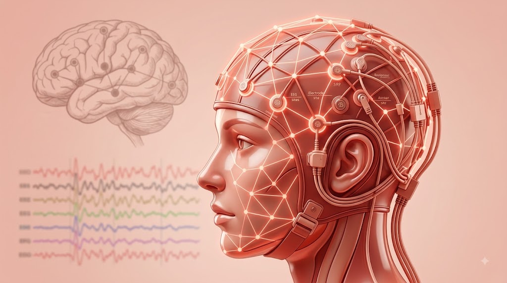
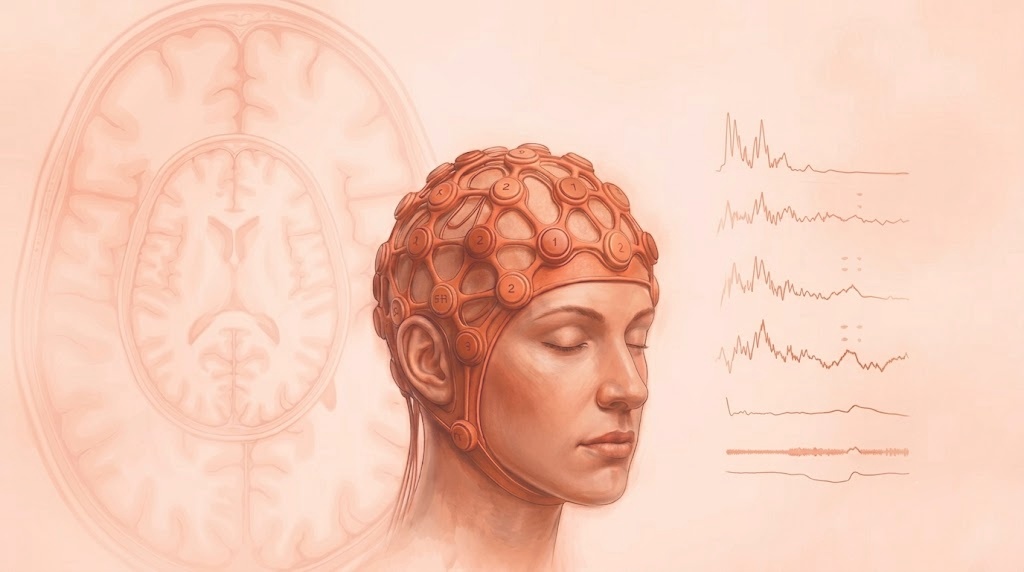
Your brain's movement-control center gives off a steady electrical rhythm when it's at rest. The moment you prepare to move, actually move, or even just imagine moving, that rhythm dips; a change researchers call desynchronization. The dip is strongest on the side of the brain opposite the body part involved: imagining a left-hand movement causes the biggest dip on the right side, and vice versa. That's the exact pattern every motor-imagery brain-computer interface is trained to recognize.
Recent research has sharpened this picture. A 2025 study found that imagining a movement and imagining a touch produce a similar brain-wave dip, but light up blood flow in different brain regions: meaning a rehab device needs to check it's picking up movement-related activity specifically, not something that just looks similar on the surface. Another 2025 study looked at why some people are better at motor imagery than others, and found brain-wave patterns that predict who's likely to succeed at it; which may help explain why some stroke patients respond faster to this training than others.
A third study found this same rhythm, measured on the same side of the brain as the imagined limb rather than the opposite side, connects to a different nerve pathway tied to shoulder movement; a hint that this approach could expand beyond hand rehab.
Real-world examples
This isn't just theory. A large clinical study already established that stroke patients can reliably learn to use this brain signal to control a device. More recent work has mapped how imagining a movement changes activity across brain regions working together, not just in one spot, and reviewed how this signal changes across a person's lifespan, relevant, since most stroke patients are older adults.
Where this is heading
Research is moving from a simple left-versus-right signal toward a much more detailed picture: which exact brain-wave pattern, on which side of the brain, predicts recovery for which body part. That detail is what will let future rehab devices go beyond hand movement to shoulders, ankles, and other joints affected by stroke.
Know More
[1] What Is the Mu Rhythm in EEG?
[2] Mu and beta rhythm topographies during motor imagery and actual movements.
[6] Motor Imagery Classification Using Mu and Beta Rhythms of EEG.
[7] Motor-Related Mu/Beta Rhythm in Older Adults: A Comprehensive Review.
Further reading




Subscribe to Neurotech Pulse
A roundup of the latest in neurotech covering breakthroughs, products, trials, funding, approvals, and industry trends straight to your inbox.