BCI Kickstarter #02: Fundamentals of Neuroscience for BCI
Welcome back to our BCI crash course! In the previous blog, we explored the basics of BCIs and different approaches to decoding brain signals. Today, we will explore the fascinating world of neuroscience to understand the foundation upon which these incredible technologies are built. This blog will focus on the electrical activity of the brain, particularly relevant for EEG-based BCIs. By understanding how neurons communicate and generate the rhythmic oscillations that EEG measures, we can gain valuable insights into the development and application of BCI systems.

Basic Brain Anatomy and Function: Your Brain's Control Center
The brain, the most complex organ in the human body, is the command center for our thoughts, emotions, and actions. To understand how BCIs tap into this intricate network, let's explore some key anatomical structures and their functions.
Brain Divisions: A Three-Part Harmony
The brain is broadly divided into three main sections:
- Forebrain: The largest and most evolved part of the brain, the forebrain is responsible for higher-level cognitive functions like language, reasoning, and problem-solving. It also processes sensory information from our environment, controls voluntary movement, and regulates emotions and motivations.
- Midbrain: Situated between the forebrain and hindbrain, the midbrain plays a crucial role in relaying sensory information to higher brain centers. It's also involved in motor control, particularly for eye movements, and in regulating sleep-wake cycles and arousal.
- Hindbrain: The oldest and most primitive part of the brain, the hindbrain is responsible for controlling vital autonomic functions such as breathing, heart rate, and blood pressure. It also coordinates balance and movement.

For BCI applications, the forebrain, particularly the cerebrum, is of primary interest. This is where conscious thought, decision-making, and voluntary actions originate.
Cerebral Cortex: The Brain's Outer Layer
The cerebrum's outer layer, the cerebral cortex, is a wrinkled sheet of neural tissue responsible for many of our higher cognitive abilities. It's divided into four lobes, each with specialized functions:
- Frontal Lobe: The "executive center" of the brain, the frontal lobe is responsible for planning, decision-making, working memory, and voluntary movement. It plays a crucial role in higher-level cognitive functions like reasoning, problem-solving, and language production. Damage to the frontal lobe can impair these functions and lead to changes in personality and behavior.
- Parietal Lobe: The parietal lobe processes sensory information related to touch, temperature, pain, and spatial awareness. It also integrates sensory input from different modalities, helping us form a coherent perception of our surroundings. Damage to the parietal lobe can cause difficulties with spatial navigation, object recognition, and body awareness.
- Temporal Lobe: The temporal lobe is involved in auditory processing, language comprehension, and memory formation. It contains structures like the hippocampus, crucial for long-term memory, and the amygdala, involved in processing emotions, particularly fear and aggression. Damage to the temporal lobe can impair memory, language comprehension, and emotional processing.
- Occipital Lobe: Located at the back of the brain, the occipital lobe is dedicated to visual processing. It receives input from the eyes and interprets visual information, allowing us to perceive shapes, colors, and motion. Damage to the occipital lobe can lead to visual impairments, including blindness or difficulty recognizing objects.

Gray and White Matter: The Brain's Building Blocks
The brain is composed of two main types of tissue:
- Gray Matter: Gray matter gets its color from the densely packed cell bodies of neurons. It is primarily involved in processing information, making decisions, and controlling actions. Gray matter is found in the cerebral cortex, basal ganglia, thalamus, and other brain regions involved in higher-level cognitive functions.
- White Matter: White matter is composed of myelinated axons, the long, slender projections of neurons that transmit electrical signals. Myelin, a fatty substance, acts as an insulator, allowing signals to travel faster and more efficiently. White matter forms the "wiring" that connects different brain regions, enabling communication and coordination between them.
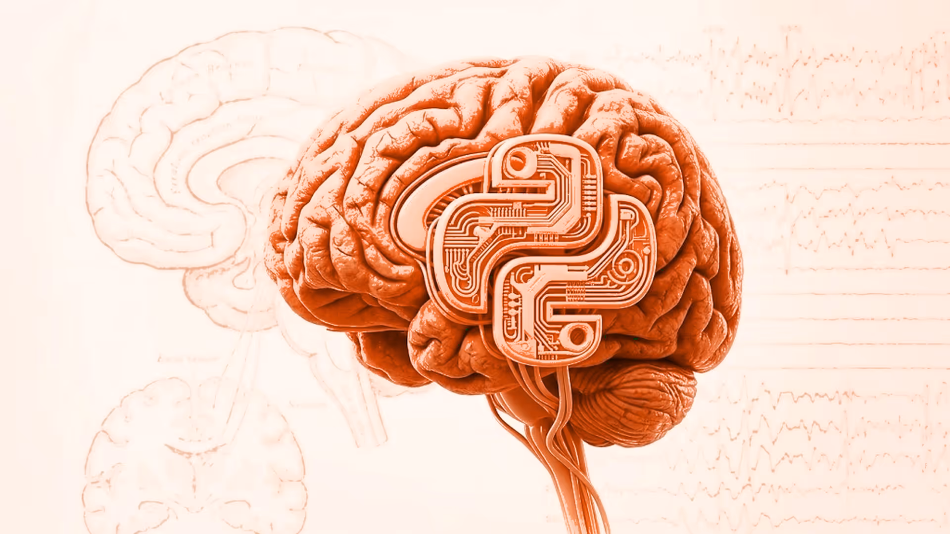
Neural Signaling and Brain Rhythms: The Electrical Symphony of Your Brain
To understand how EEG-based BCIs work, we need to dive deeper into how neurons communicate and generate the electrical signals that EEG measures. This intricate process involves a complex interplay of electrical impulses, chemical messengers, and rhythmic oscillations.
Neurons and Synapses: The Building Blocks of Communication
Neurons are specialized cells that transmit information throughout the nervous system. They have a unique structure:
- Dendrites: Branch-like extensions that receive signals from other neurons.
- Cell Body (Soma): Contains the nucleus and other cellular machinery.
- Axon: A long, slender fiber that transmits electrical signals away from the cell body.
- Synapse: A small gap between the axon of one neuron and the dendrite of another, where communication occurs.
Electrical Signaling: The Language of Neurons
Neurons communicate using electrical impulses called action potentials. These brief, rapid changes in electrical charge travel down the axon, triggered by a complex interplay of ion channels that regulate the flow of charged particles across the neuron's membrane.
Think of an action potential like a wave traveling down a rope. It's an all-or-nothing event; once triggered, it propagates down the axon at a constant speed and amplitude.
When an action potential reaches the synapse, it triggers the release of neurotransmitters, chemical messengers that cross the synaptic gap and bind to receptors on the receiving neuron. This binding can either excite or inhibit the receiving neuron, modulating its likelihood of firing its own action potential.
Neurotransmitters and Receptors: Fine-Tuning the Signals
Neurotransmitters are the brain's chemical messengers, playing a crucial role in regulating mood, cognition, and behavior. Here are some key neurotransmitters relevant to BCI applications:
- Glutamate: The primary excitatory neurotransmitter in the brain, involved in learning, memory, and synaptic plasticity.
- GABA (Gamma-Aminobutyric Acid): The primary inhibitory neurotransmitter, important for calming neural activity and preventing overexcitation.
- Dopamine: Involved in reward, motivation, and motor control, playing a crucial role in Parkinson's disease.
- Acetylcholine: Plays a vital role in muscle contraction, memory, and attention.
Each neurotransmitter binds to specific receptors on the receiving neuron, triggering a cascade of intracellular events that ultimately modulate the neuron's electrical activity.
EEG Rhythms and Oscillations: Decoding the Brain's Rhythms
EEG measures the synchronized electrical activity of large groups of neurons firing together, generating rhythmic oscillations that reflect different brain states. These oscillations are categorized into frequency bands:
- Delta (1-4 Hz): The slowest brainwaves, dominant during deep sleep and associated with memory consolidation.
- Theta (4-8 Hz): Prominent during drowsiness, meditation, and creative states, often linked to cognitive processing and working memory.
- Alpha (8-12 Hz): Associated with relaxed wakefulness, particularly with eyes closed. Alpha waves are suppressed during mental exertion and visual processing.
- Beta (12-30 Hz): Reflect active thinking, focus, and alertness. Increased beta activity is observed during tasks requiring sustained attention and cognitive effort.
- Gamma (30-100 Hz): The fastest brainwaves, associated with higher cognitive functions, sensory binding, and conscious awareness.
By analyzing these rhythmic patterns, EEG-based BCIs can decode user intent, mental states, and even diagnose neurological conditions.
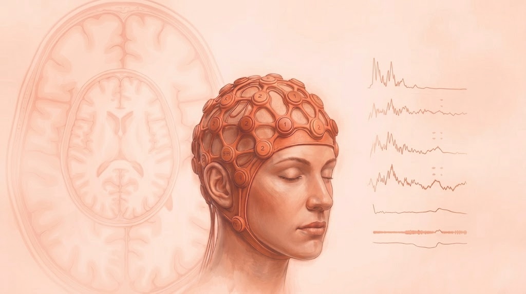
Electroencephalography (EEG) and its Significance in BCI: Capturing the Brain's Electrical Whispers
EEG, as we've mentioned throughout this post, is a powerful tool for capturing the electrical activity of the brain, making it a cornerstone of many BCI systems. Let's explore how EEG works and why it's so valuable for decoding brain signals.
How EEG Works: Recording the Brain's Electrical Symphony
EEG measures the electrical potentials generated by synchronized neuronal activity in the cerebral cortex. This is achieved using electrodes placed on the scalp, which detect the tiny voltage fluctuations produced by these electrical currents.
The electrodes are typically arranged according to the 10-20 system, a standardized placement system that ensures consistent and comparable recordings across different individuals and research studies.
The 10-20 System: A Standardized Map for EEG Recording
The 10-20 system is the internationally standardized method for placing EEG electrodes. It provides a consistent framework for recording and interpreting EEG data, allowing researchers and clinicians worldwide to communicate and compare results effectively.
The system is based on specific anatomical landmarks on the skull:
- Nasion: The indentation at the top of the nose, between the eyebrows.
- Inion: The bony prominence at the back of the head.
- Preauricular Points: The depressions just in front of each ear.
Electrodes are placed at intervals of 10% or 20% of the total distance between these reference points, forming a grid-like pattern that covers the scalp.
Each electrode is labeled with a letter and a number:
- Letters: Represent the underlying brain region (Fp for prefrontal, F for frontal, C for central, P for parietal, T for temporal, O for occipital).
- Numbers: Indicate the hemisphere (odd numbers for the left, even numbers for the right, and z for midline).
This standardized system ensures that electrodes are consistently placed in the same locations across different individuals, facilitating reliable comparisons and analysis of EEG data.
High-Density vs. Low-Density Systems: A Matter of Resolution
EEG systems vary in the number of electrodes they use, ranging from a few electrodes in consumer-grade headsets to hundreds of electrodes in research-grade systems.
- High-Density Systems: Provide higher spatial resolution, allowing for more precise localization of brain activity. They are commonly used in research settings for investigating complex cognitive processes and mapping brain function.
- Low-Density Systems: Offer portability and affordability, making them suitable for consumer applications like neurofeedback, meditation training, and sleep monitoring. However, their lower spatial resolution limits their ability to pinpoint specific brain regions.
The choice of system depends on the specific application and the desired level of detail in capturing brain activity.
Types of EEG Electrodes: From Wet to Dry
Various types of EEG electrodes are available, each with its own advantages and disadvantages:
- Wet Electrodes: Require a conductive gel or paste to enhance electrical contact with the scalp. They generally provide better signal quality but can be more time-consuming to apply.
- Dry Electrodes: Don't require conductive gel, making them more convenient and user-friendly, but they might have slightly lower signal quality.
EEG Montages: Choosing Your Viewpoint
EEG montages refer to the way electrode pairs are connected to create the electrical signals displayed. Different montages highlight different aspects of brain activity and can influence the interpretation of EEG data.
Common montages include:
- Bipolar Montage: Each channel represents the voltage difference between two adjacent electrodes, emphasizing localized activity and minimizing the influence of distant sources.
- Referential Montage: Each channel represents the voltage difference between an active electrode and a common reference electrode (e.g., linked mastoids, average reference). This montage provides a broader view of brain activity across regions but can be more susceptible to artifacts from the reference electrode.
The choice of montage depends on the research question or BCI application. Bipolar montages are often preferred for studying localized brain activity, while referential montages are useful for examining activity across broader brain regions.
Further Reading and Resources:
- Principles of Neural Science – Kandel et al
- HarvardX: Fundamentals of Neuroscience, Part 1: The Electrical Properties of the Neuron (https://www.edx.org/learn/neuroscience/harvard-university-fundamentals-of-neuroscience-part-1-the-electrical-properties-of-the-neuron)
- HarvardX: Fundamentals of Neuroscience, Part 2: Neurons and Networks (https://www.edx.org/learn/neuroscience/harvard-university-fundamentals-of-neuroscience-part-2-neurons-and-networks)
- HarvardX: Fundamentals of Neuroscience, Part 3: The Brain (https://www.edx.org/learn/neuroscience/harvard-university-fundamentals-of-neuroscience-part-3-the-brain)
Ready to Dive Deeper into EEG Signal Processing?
This concludes our exploration of the fundamentals of neuroscience for BCI. In the next post, we'll dive into the practical aspects of EEG signal acquisition and processing, exploring the techniques used to extract meaningful information from the raw EEG data.
Stay tuned for our next BCI adventure!
Further reading


Subscribe to Neurotech Pulse
A roundup of the latest in neurotech covering breakthroughs, products, trials, funding, approvals, and industry trends straight to your inbox.